| biology_3201_outline.pdf |
| objectives_unit_1_nervous_system.docx |
Unit 1: Maintaining Dynamic Equilibrium II (The Nervous System)
Introduction:
- Cells, tissues, organs, and ultimately organisms must maintain a biological balance despite changing external conditions.
- Homeostasis is the state of internal balance so critical to existence.
- It represents a dynamic equilibrium displaying constant interactions and checks and balances both within organisms and between organisms and their environment.
- There are a variety of systems within living things responsible for the maintenance of this delicate balance.
- This unit introduces the role played by the nervous (electrochemical) and endocrine (chemical) systems in humans.
The Nervous System (p. 390-419)
CNS - Central Nervous System (brain and spinal cord)
PNS - Peripheral Nervous System (nerves coming off brain and spinal cord)
Objective #1 & 2
The Central Nervous System
- receives sensory information and initiates motor control
- made up of the brain and spinal cord
- is covered by 3 protective membranes called meninges. (P.399)
- between the inner two layers is a fluid called cerebrospinalfluid (p.393 & 407) that protects the CNS from injury and collects and drains away waste products.
- the skull (cranium) also aids in the protection of the brain
Example from class (rock in a water balloon in a box) - brain in cerebriospinal fluid, surrounded by meninges, inside the skull.
Objective #3
The Peripheral Nervous System (PNS) (p.393) - all nerves outside the brain and spinal cord
PNS - Divided into Somatic Nervous System & Autonomic Nervous System
Somatic Nervous System
- nerves connecting body parts under, mostly, voluntar (conscious) control, eg. walking, flexing biceps. Some of the nerves are sensory, carrying impulses from the body=s sense organs to the CNS and some are motor nerves that carry impulses from the CNS to the muscles.
- reflex actions are somatic but do not require a conscious decision. (Exception!)
Autonomic Nervous System
- nerves connecting body parts that have involuntary (unconscious) action, eg. Stomach muscles contract to digest food.
- affects glands, smooth muscle (digestion) and cardiac muscles (heart).
Autonomic is further divided into Sympathetic and Parasympathetic Systems
Sympathetic:
- sets off a 'fight or flight' reaction, that prepares the body to deal with an immediate threat. Heart rate and breathing rate increase. Blood sugar is released from the liver for energy. Ganglia located near spine.
Parasympathetic
- has opposite effect. When the threat has passed, the nerves of this system slow heart rate and breathing rate and reverse the effects of the sympathetic nervous system. Ganglia located near the organs they affect.
(See Figure 12.5, page 394, for effects of these systems.)
Objective #4
The nervous system is responsible for receiving info from internal and external stimuli and quickly responding to that info.
For a nervous response to occur the following requirements are
necessary: (PLA)
1. sensoryreceptors to detect a stimulus (skin, eye, ear)
2. method for impulse transmission (neurons)
3. interpretation and analysis of impulses (brain, spinal cord)
4. response carried out by an effector (muscle, gland)
Refer to reflec arc on page 396. We drew the diagram in our notes and added the underlined words to the diagram.
Objective #5
reflect arc - the nerve pathway that leads from the stimulus to the reflex action.
Objective #6
Obtain handout from class. Label diagram below (p. 395 for help) of neuron
1. cell body
2. nucleus
3. dendrite
4. axon
5. schwann cell nucleus (N/A for evaluation)
6. myelin sheath
7. node of Ranvier
8. schwann cell
9. terminal branches
10. synaptic knob
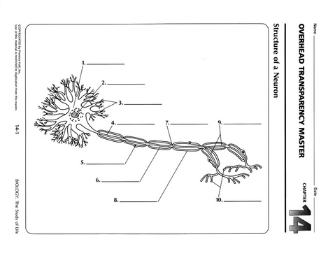
The Neuron (p.395)
a nerve cell -> the structural and functional unit of the nervous system. Carries info from one location in the body to another. Capable of surviving over 100 yrs., since most do not undergo cell division after adolescence.
Parts of a neuron: (Refer to Figure: 12.6, p. 395)
dendrites- site for receiving signals from other neuron. # can range from 1- 1000's, depending on function.
cell body - has large centrally located nucleus with large
nucleolus. Contains variety of cell structures (mitochondria, lysosomes, Golgi complex, ER)
axon - long, cylindrical extension of the cell body, ranging 1mm-1m.
Axon terminals are distal terminations of the branches of an axon.
myelin sheath -> fatty layer covering axon; insulates the neuron,to prevent short circuiting of impulse to neighbouring neurons. This
allows several neurons to exist side by side within a nerve (as found in PNS).
Schwann cells -> insulating cells that together form the
myelin sheath; also may allow regeneration of damaged neuron, if damage is not
severe.
node of Ranvier -> the gap between each Schwann cell; a
nerve impulse travelling along a myelinated neuron is able to jump from node to
node, speeding up the impulse wave.
Objective #7
Classes of Neurons: (p.395)
sensory neurons -> pick up sensory info from the environment via sensory receptors (eg. temperature and pressure receptors in the
skin) and carry it to the CNS.
interneurons(association ) -> carries info from one neuron to another; can receive info from sensory neuron or another interneuron.
motor neurons -> carries info from CNS to an effector ie. a muscle fiber (to contract) or to a gland (to secrete a substance).
eg. sensory neuron —> interneuron —> motor neuron
(in skin) (spinal cord) (muscles)
The three major types of neurons can be illustrated in a simple REFLEX ARC:
( see page 395 and Figure 12.7 p. 396)
1. sensory receptors (dendrites) pick up stimulus and bring it to the spinal cord.
2. within spinal cord, the impulse is passed to the interneuron which passes info to:
a. a motor neuron that transmits the impulse to a muscle (effector)
and
b. to other interneurons that transmit the impulse to the brain,making it aware of what has just happened.
In a reflex, the effector reacts before the brain interprets the message (awareness occurs).
eg. your hand pulls away from a hot stove before you feel the pain! (screaming “Owww!” comes after even that!)
Objective #8
Biology 3201
Name: _________________________________
Exploring the Brain
Read pages 397-98 in your textbook
1. How did the study of patients with brain damage or injury lead early researchers to discoveries of the functions of the parts of the brain?
· Damage – loss of functios/changes in behaviour
· Abnormalities of the structure of the brain ‘must have’ caused these problems, thought earlier scientists
· The damaged/abnormal area controls the function (walking, talking, etc) that was changed by the damage or disorder
2. What is an EEG machine (what does EEG stand for)? Electroencephalograph
3. What does an EEG machine measure and how is it used for brain study?
· Measures electrical activity (electrodes placed in certain regions)
· Used to diagnose epilepsy, detect tumors, diagnose sleep problems.
4. How has direct stimulation of the brain tissue been used to ‘map’ the functioning of various brain parts?
· Direct stimulation is able to occur because there are no pain receptors in the brain (no pain felt)
· When the temporal lobe was stimulated, the person ‘heard voices’ of family members who weren’t there, or heard music from a rock concert attended in the past.
· By stimulating the different lobes, the scientists could determine which lobes are linked to certain functions (in this case, they linked the temporal lobe to hearing)
CAT scans
· Cross sectional view of body using x-rays
· Creates a 3-d computer image of the brain to reveal things like tumors
PET scans
· A person is injected with a radioactive substance which releases positrons. Positrons are released by activated areas of the brain (i.e., a person ‘looking’ has high positron activity at the back of the brain
· Used for many disorders (brain tumors, seizures)
MRI scans
· Uses an electromagnet (magnetic field) to cause the body to emit radio waves which causes a computer generated image that shows vital organs, such as the brain, and identifies abnormalities such as tumors.
Objective #9 Parts of the brain: (p.399-400)
(i) the cerebrum
- divided into left and right hemispheres, by a dividing line called the fissure.
- left hemisphere controls movement in the right side of the body and the right hemisphere controls movement of the left side of the body. (Left is usually dominant over right)
- controls reasoning (thinking), memory and voluntary (under your control) muscle action.
- where intelligent thought occurs (center of human consciousness)
- sensory information and emotions are interpreted
- the cerebrum is divided into 4 lobes:(p.400)
(a) frontal lobe -> (intelligence, reasoning, control of striated muscles.)
(b) temporal lobe -> (receives info from ears for hearing.)
(c) occipital lobe -> (receives info from eyes for vision.)
(d) parietal lobe -> (sensory info from skin and skeletal muscles and associated with sense of taste.)
(ii) cerebellum
- located at the base of the brain (old woman's bun)
- helps in balance and muscle coordination to produce smooth movement, eg. walking, ballet, throwing a ball, etc.
- makes up only 10% of brain volume (space), but 50% of the brain=s neurons (nerves).
(iii) medulla oblongata
- located just above the spinal cord
- has a number of functions, each related to a particular structure:
a cardiac centre controls heart rate and the force of the heart=s contractions,
b. vasomotor centre adjusts blood pressure by controlling the diameter of blood vessels,
c. respiratory centre controls the rate and depth of breathing,
d. reflex centres for vomiting, couphing, hiccuping, and swallowing.
- any damage to this part of the brain is usually fatal.
(iv) thalamus
- relay station, directing sensory information to the appropriate part of the brain (cerebrum if minor, hypothalamus if major)
- it receives sensations of touch, pain, heat, and cold as well as info from the muscles.
- screens out info that does not have priority; eg. focussing on a conversation with one person in a room where many are talking.
(v) hypothalamus
- main control centre for the autonomic nervous system.
- controls many bodily activities that maintain homeostasis; eg. hunger, thirst, temperature regulation, blood pressure, water balance.
- enables the body to respond to external threats by sending impulses to various internal organs via the sympathetic nervous system and re-establishes homeostasis after the threat by stimulating the parasympathetic nerves.
(vi) corpus callosum
- joins the two hemispheres of the brain, transferring impulses from one hemisphere to the other.
- a layer of white matter, made up of axons.
(vii) midbrain
- segment of brainstem between cerebrum and pons
- functions in sight and hearing
(viii) pons -
- bulb-like structure of the brainstem
- sends signals between cerebellum and CNS
- functions with medulla to regulate breathing
- reflex centres involved in head movement
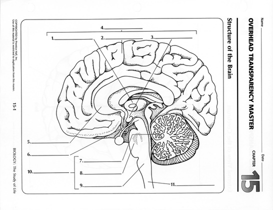
Brain structures - diagram
1. thalamus
2. cerebrum
3. hypothalamus
4. forebrain **
5. corpus callosum
6. midbrain
7. cerebellum
8. pons
9. medulla oblongata
10. hindbrain **
11. spinal cord
Objective 10, 11, &13 below in powerpoint
a nerve cell -> the structural and functional unit of the nervous system. Carries info from one location in the body to another. Capable of surviving over 100 yrs., since most do not undergo cell division after adolescence.
Parts of a neuron: (Refer to Figure: 12.6, p. 395)
dendrites- site for receiving signals from other neuron. # can range from 1- 1000's, depending on function.
cell body - has large centrally located nucleus with large
nucleolus. Contains variety of cell structures (mitochondria, lysosomes, Golgi complex, ER)
axon - long, cylindrical extension of the cell body, ranging 1mm-1m.
Axon terminals are distal terminations of the branches of an axon.
myelin sheath -> fatty layer covering axon; insulates the neuron,to prevent short circuiting of impulse to neighbouring neurons. This
allows several neurons to exist side by side within a nerve (as found in PNS).
Schwann cells -> insulating cells that together form the
myelin sheath; also may allow regeneration of damaged neuron, if damage is not
severe.
node of Ranvier -> the gap between each Schwann cell; a
nerve impulse travelling along a myelinated neuron is able to jump from node to
node, speeding up the impulse wave.
Objective #7
Classes of Neurons: (p.395)
sensory neurons -> pick up sensory info from the environment via sensory receptors (eg. temperature and pressure receptors in the
skin) and carry it to the CNS.
interneurons(association ) -> carries info from one neuron to another; can receive info from sensory neuron or another interneuron.
motor neurons -> carries info from CNS to an effector ie. a muscle fiber (to contract) or to a gland (to secrete a substance).
eg. sensory neuron —> interneuron —> motor neuron
(in skin) (spinal cord) (muscles)
The three major types of neurons can be illustrated in a simple REFLEX ARC:
( see page 395 and Figure 12.7 p. 396)
1. sensory receptors (dendrites) pick up stimulus and bring it to the spinal cord.
2. within spinal cord, the impulse is passed to the interneuron which passes info to:
a. a motor neuron that transmits the impulse to a muscle (effector)
and
b. to other interneurons that transmit the impulse to the brain,making it aware of what has just happened.
In a reflex, the effector reacts before the brain interprets the message (awareness occurs).
eg. your hand pulls away from a hot stove before you feel the pain! (screaming “Owww!” comes after even that!)
Objective #8
Biology 3201
Name: _________________________________
Exploring the Brain
Read pages 397-98 in your textbook
1. How did the study of patients with brain damage or injury lead early researchers to discoveries of the functions of the parts of the brain?
· Damage – loss of functios/changes in behaviour
· Abnormalities of the structure of the brain ‘must have’ caused these problems, thought earlier scientists
· The damaged/abnormal area controls the function (walking, talking, etc) that was changed by the damage or disorder
2. What is an EEG machine (what does EEG stand for)? Electroencephalograph
3. What does an EEG machine measure and how is it used for brain study?
· Measures electrical activity (electrodes placed in certain regions)
· Used to diagnose epilepsy, detect tumors, diagnose sleep problems.
4. How has direct stimulation of the brain tissue been used to ‘map’ the functioning of various brain parts?
· Direct stimulation is able to occur because there are no pain receptors in the brain (no pain felt)
· When the temporal lobe was stimulated, the person ‘heard voices’ of family members who weren’t there, or heard music from a rock concert attended in the past.
· By stimulating the different lobes, the scientists could determine which lobes are linked to certain functions (in this case, they linked the temporal lobe to hearing)
CAT scans
· Cross sectional view of body using x-rays
· Creates a 3-d computer image of the brain to reveal things like tumors
PET scans
· A person is injected with a radioactive substance which releases positrons. Positrons are released by activated areas of the brain (i.e., a person ‘looking’ has high positron activity at the back of the brain
· Used for many disorders (brain tumors, seizures)
MRI scans
· Uses an electromagnet (magnetic field) to cause the body to emit radio waves which causes a computer generated image that shows vital organs, such as the brain, and identifies abnormalities such as tumors.
Objective #9 Parts of the brain: (p.399-400)
(i) the cerebrum
- divided into left and right hemispheres, by a dividing line called the fissure.
- left hemisphere controls movement in the right side of the body and the right hemisphere controls movement of the left side of the body. (Left is usually dominant over right)
- controls reasoning (thinking), memory and voluntary (under your control) muscle action.
- where intelligent thought occurs (center of human consciousness)
- sensory information and emotions are interpreted
- the cerebrum is divided into 4 lobes:(p.400)
(a) frontal lobe -> (intelligence, reasoning, control of striated muscles.)
(b) temporal lobe -> (receives info from ears for hearing.)
(c) occipital lobe -> (receives info from eyes for vision.)
(d) parietal lobe -> (sensory info from skin and skeletal muscles and associated with sense of taste.)
(ii) cerebellum
- located at the base of the brain (old woman's bun)
- helps in balance and muscle coordination to produce smooth movement, eg. walking, ballet, throwing a ball, etc.
- makes up only 10% of brain volume (space), but 50% of the brain=s neurons (nerves).
(iii) medulla oblongata
- located just above the spinal cord
- has a number of functions, each related to a particular structure:
a cardiac centre controls heart rate and the force of the heart=s contractions,
b. vasomotor centre adjusts blood pressure by controlling the diameter of blood vessels,
c. respiratory centre controls the rate and depth of breathing,
d. reflex centres for vomiting, couphing, hiccuping, and swallowing.
- any damage to this part of the brain is usually fatal.
(iv) thalamus
- relay station, directing sensory information to the appropriate part of the brain (cerebrum if minor, hypothalamus if major)
- it receives sensations of touch, pain, heat, and cold as well as info from the muscles.
- screens out info that does not have priority; eg. focussing on a conversation with one person in a room where many are talking.
(v) hypothalamus
- main control centre for the autonomic nervous system.
- controls many bodily activities that maintain homeostasis; eg. hunger, thirst, temperature regulation, blood pressure, water balance.
- enables the body to respond to external threats by sending impulses to various internal organs via the sympathetic nervous system and re-establishes homeostasis after the threat by stimulating the parasympathetic nerves.
(vi) corpus callosum
- joins the two hemispheres of the brain, transferring impulses from one hemisphere to the other.
- a layer of white matter, made up of axons.
(vii) midbrain
- segment of brainstem between cerebrum and pons
- functions in sight and hearing
(viii) pons -
- bulb-like structure of the brainstem
- sends signals between cerebellum and CNS
- functions with medulla to regulate breathing
- reflex centres involved in head movement
Brain structures - diagram
1. thalamus
2. cerebrum
3. hypothalamus
4. forebrain **
5. corpus callosum
6. midbrain
7. cerebellum
8. pons
9. medulla oblongata
10. hindbrain **
11. spinal cord
Objective 10, 11, &13 below in powerpoint
| nerve_impulse.pptx |
Objective #12 – Brain and spinal cord injuries
Damage of the CNS
• If neurons in the CNS are damaged, they cannot regenerate
• However, if a part of the brain is damaged, it’s function may be taken over by another part of the brain (This ‘rerouting’ often takes extensive rehabilitation)
• Damage to the spinal cord is usually permanent (paralysis below point of injury)
Stroke
• A stroke occurs when a portion of the brain does not receive enough oxygen and dies (often due to blood clot or cholesterol plaque).
• This occurs when there is a disruption in the blood supply to the brain.
• If the disruption is not restored, the brain tissue that is deprived of oxygen will be permanently damaged (full blown stroke)
Treatment for stroke
• Clot busting drugs
• Medicine must be taken within a short time of the onset of the stroke (within 3 hours)
• Aspirin may be prescribed to those showing symptoms of stroke (reduces stickiness of platelets, decreasing the chance of a clot occurring)
Risk of taking aspirin to prevent stroke
• Strokes can also be caused by aneurysms (blood vessel breaks in the brain)
• If someone is taking aspirin in this case, it can induce more bleeding
Spinal Cord injuries
• Spinal cord injuries can vary, from having no effect on the patient to a "complete" injury which means a total loss of function.
• Treatment: starts with restraining the spine and controlling inflammation but treatment can vary
• Can require substantial physical therapy and rehabilitation
Brain and spinal repair research
• Research: on mice for spinal cord repair
• Also in cancer patients – using a drug they detected the formation of new brain cells
• These new neurons are derived from stem cells in some areas of the brain
Objective 14 & 15
Effects of Neurotransmitters:
•acetycholine
- primary neurotransmitter for somatic and parasymp. NS=s
- can be excitatory -> skeletal muscles or
- can be inhibitory -> cardiac muscle
Note: The neurotransmitter released into a synapse and attached to the postsynaptic receptors is immediately broken down by an enzyme released from the presynaptic neuron; cholinesterase breaks down acetylcholine. (Objective 15)
•noradrenaline (norepinephrine)
- primary transmitter for sympathetic NS
- regulates alertness, sleep-wakefulness cycle, associated with memory, attention focus (eliminating distractions), and learning.
•glutamate
- neurotransmitter for cerebral cortex (cerebrum) – memory and learning
- accounts for 75% of excitatory transmissions in the brain
- low glutamate – agitation, memory loss, sleep problems
•gamma aminobutyric acid (GABA)
- most common inhibitory neurotransmitter in the brain
- GABA binds to receptors and results in chloride being allowed into the neuron or it allows more flow of potassium ions out of the cell/axon (raising the threshold)
- a lack of GABA can result in seizures, anxiety disorders, and muscle tremors
•dopamine
- elevates mood and contros skeletal muscles
- acts to carry messages between areas of the brain controlling body movements
- lack of dopamine – Parkinson’s
•seratonin
- involved in alertness, sleepiness, thermoregulation, mood, appetite, memory/learning
- lack of serotonin – depression, hot flashes, sadness/crying, loss of appetite or cravings
Some drugs have been developed to stimulate or inhibit specific Neurotransmitters:
•eg. valium -> stimulates GABA (alleviating anxiety)
•Prozac -> antidepressant that enhances the action of serotonin
http://www.holisticouncil.org/neuro.html - info on neurotransmitters
| biology_3201_all_stse_readings.pdf |
